• German
  • English
  • Warenkorb
  • Login/Benutzerkonto
Verlag Dr. Friedrich Pfeil
  • [avia_search]
  • Shop
    • Login/Benutzerkonto
  • Gesamtverzeichnis
  • Archäologie
    • Bücher
    • Reihen & Zeitschriften
    • Blickpunkt Archäologie
  • Biologie
    • Bücher
    • Reihen & Zeitschriften
    • Ichthyological Exploration of Freshwaters
    • Meiofauna Marina
  • Geographie
    • Bücher
    • Reihen & Zeitschriften
  • Geologie
    • Bücher
    • Kinderbücher Geologie
    • Reihen & Zeitschriften
    • Wanderungen in die Erdgeschichte
    • Münchner Geowissenschaftliche Abhandlungen Reihe B
  • Ökologie
  • Paläontologie
    • Bücher
    • Reihen & Zeitschriften
    • Münchner Geowissenschaftliche Abhandlungen Reihe A
    • Handbook of Paleoichthyology
  • Sonstiges
    • Philosophie
    • Romane
    • Volkskunde
  • E-Book (PDF) Bestellungen
  • Verlag
  • Buchhandel
  • Für Autoren
  • Kontakt
  • Menü Menü
  • Einkaufswagen Einkaufswagen
    0Einkaufswagen
  • Link zu Facebook
  • Link zu X

Suche

Search Search
Blick ins Buch:
SOLOUNIAS, Nikos

The Horse – Head, Dentition and Limbs

Anatomy and Evolution

2024. [Englisch] – 132 Seiten, 89 Farbabbildungen, 10 Schwarzweißabbildungen.
30,3 × 21,3 cm, Hardcover.

ISBN: 978-3-89937-280-7
Artikelnummer: 9783899372807 Kategorien: Biologie, Bücher Biologie, Bücher Paläontologie, Paläontologie

78,00 €

zzgl. Versandkosten / Versandkostenfrei in D


  • Beschreibung
  • Inhalt
  • Preface
  • Vorwort
  • Autor
  • Faltblatt
  • Kundenmeinungen
  • Rezensionsexemplar

Nikos Solounias ist deeply involved with horses and their ancient relatives of times long past. For several decades he ventured out into the world to learn everything he can about the history of the equids. His approach of in-depth examination of the modern horse and constant comparison with paleontologic findings afford him rare insights into anatomy and function. In this book the focus is set on three remarkable discoveries in the head, the dentition and the limbs of the modern horse.

In the equine head the the interaction of nasal cavity, the nostrils but especially of the pre­orbital fossa and the nasal diverticulum are examined in detail. This not only allows new insights into the abilities and functions of the nose but also gives insights into the evolution of the anatomy of the equid face.

In the case of the dentition the interaction between the teeth and the vegetation while chewing provides new insights that explain the structure of the teeth. Additionally new theories about hypsodonty give further insight into the anatomy and evolution of the teeth.

His research on the lower limbs of the horse provide the perhaps most striking results. Investigations of embryos andpaelontologic finds support the novel five-digit scenario of the meta­carpals and metatarsals. This reveals misunderstandings and new ideas about monodactyly.

Contents 5
Preface 6
Introduction 7

The nasal cavity and the anatomy of the preorbital area 9
The nasal cavity 13
The preorbital fossa of the horse (POF) 24
The air sacs of the horse 34
Paleontology 37
Merychippus insignis 38
Merchippus (Lakobatipus) sp.42
Cremahipparion proboscideum  47
Embryology 56
Comparative species 64
The lowland tapir – Tapirus terrestris and Tapirus indicus Malayan tapir  64
The white-tailed deer – Odocoileus virginianus 67
Sound production in the Cetacea and similarities to the horse  69
The Indian elephant – Elephas maximus 70
The cotton tail rabbit – Sylvilagussp. 72
Probable developmental functions for the preorbital fossae 74
Conclusion 79

The dentition – mastication and tooth wear 85
A short introduction to horse dentition 85
The anatomy of the Equus hypsodont teeth 89
Mastication and the anatomy of tooth wear 98
Motion of the jaw and some forces of mastication 100
The misunderstood lower dentition – a remarkable mutation 103
Cementum wear106
Tooth mesowear in equids109

The anatomy and embryology of the forelimb and feet 111
Brief osteological anatomy114
The histology of the horse hoof116
Evolution121
The horse anatomy126
Conclusion 128

References 129
Acknowledgments 132

I have been interested in bones since I was four years old. It was a deep internal drive and I was born with it. It is my demon. I had made collections of bones, fossils and x-rays in elementary school. When I was 12-year-old, John Melentis showed me the Pikermi fossils at the museum in Athens. That day blew my mind. I was sure I was deeply connected with the Miocene fossils. Our family comes from Samos (all four grandparents) and when young my grandmother told me a story that on Samos there was an elephant cemetery where elephants go to die. In 1959 I went to find the elephant cemetery and I run into fossils at Samos. The very first fossil I found near the Mytilinii Village was a femur of Samotherium (an extinct giraffid). Other fossils were rhino bones and hipparions. I came to the United States to study biology in 1966. A cold December day was the second day of my arrival in the USA, a friend took me to the American Museum of Natural History and informed Malcolm McKenna and Thomas Rich that a young person was interested in paleontology. Rich invited me to go to Alberta to find early mammals. After volunteering there for a few days, I told them I wanted to study Pikermi and Samos. In 1966 Malcolm McKenna and Morris Skinner showed me the Samos collection at the American Museum of Natural History in New York. I am from Samos and for this reason I was encouraged to the study of Samos fossils of the museum. The Samos collection was made by the eminent paleontologist Barnum Brown in 1921-1924. The Samos collection is large and impressive. I became immediately attached to it. All these animals had lived near my home millions of years ago. Among other fossils there was a hipparion horse. It was named then Hipparion proboscideum. It was stunning to me how similar and yet how different it was from the modern horse. Overall it looked like a modern horse. But there were some features that were remarkably different. It had a deep depression in front of its eye and the hooves were not one but three. Yet, it was in most ways like the modern horse. I wondered why there was a strange depression in front of the eye and why the three hooves? What were the differences in the lips and face and what did it eat? That specimen left an impression on me and directed my career. Since then, I have studied biology and geology. I have specialized in mammals and the fossils from the island of Samos. I studied the geology of Samos with the supervision of John VanCouvering and Marc Weidmann. I have excavated for rodents at Samos. In Lausanne Marc Weidmann was my support and helped me a great deal with the Samos fossils. My wife Bernadette helped me a lot with my museum and field studies. I have studied the fossils in many natural history museums and I have tried to understand the paleoecology of the Samos animals and of the late Miocene. I have spent many years with trying to research their paleodiets and paleoecology. I have developed a tooth microwear method and a tooth mesowear method. I have excavated with the Harvard team in the Miocene Siwaliks of Pakistan. Now with this book, I am returning to the anatomy and evolution of the horse and the fascinating horse-like ancestors.

The domesticated horses are really well-known animals. There are many studies of their husbandry, anatomy, evolution and biology. The horse has been admired and loved throughout the World. Although there are several studies of its anatomy by Schmaltz (1929), Ellenberger & Baum (1894-1901), Boas & Paulli (1908) and others, the knowledge is incomplete. Certain issues about their anatomy and evolution have not been studied in detail. The present study is not a literature review. It covers new information on three parts of the horse:

1            The nasal cavity anatomy and the anatomy of the preorbital area (p. 9).

2            The dentition – mastication and tooth wear (p. 85).

3            The anatomy and embryology of the forelimb and feet (p. 111).

These areas are relevant for the understanding of horse communication, diet, and locomotion. They are a contribution in development and evolution. They relate the horse to its ancestors.
This study is inspired from the Samos Cremo­hipparion (Hipparion) proboscideum (Bernor et al. 2021). Two cranial specimens in particular stand out: the type in Bern and a female skull in Frankfurt (NHMBa C 5019 old 6; SMF 4709). These two specimens have fascinated me. They are so similar and yet so different form our modern horses.
I wanted to do something for this species that no one else has done. I did not want to measure their fossae or their teeth. I did not want to do biostrati­graphy. This study is devoted to how one can study the fossa, the diet and the anatomy of the tridactyl feet. What types of questions can be formulated and answered? How is the horse different today? What were the reasons of the observed changes?
The study focuses on new ideas, observations and hypotheses and is not meant to be an exhaustive coverage of previous research. New anatomic and functional terms are introduced in this study. The study has many figures because I believe they help understand the anatomy better than simple text. All the figures are new. I emphasize that the figures are data. The figures are many and detailed. They reflect my personality as I think in images not words. I also know that we all love figures in studies and often skip over the words.
The term domesticated horse or horse is meant to mean the domesticated horse Equus caballus. The three terms are used interchangeably throughout the text. Most of the research is based on the domesticated horse as this species was researched the most. Other species are mentioned by their scientific name. Equids refers to the taxa of the family Equidae. Equinae and Anchitheriinae are the two major subfamilies. I have selected some additional literature for further reading. I have also summarized the main points at the end of each chapter. A list of species and a list of terms in Latin are also provided at the end. In figures I use no abbreviations for the anatomical terminology. This greatly facilitates the use of the figures because it eliminates the step of having to look up what is been pointed. I use anglicized Latin [L] and Greek [G] in the text and figures as do most medical books (e. g. Gray’s Anatomy 40th edition, 2008).
During the later Cenozoic, especially in North America, there is a large adaptive radiation of extinct horses. Form the very beginning to the Oligocene there were numerous species. More than forty species were present in the Oligocene-Miocene of North America. Dinohippus is most likely the ancestor of Equus (the modern horses are all placed in one genus: Equus). Mihlbachler et al. (2011) provides a useful cladogram and the dietary evolution of equids. McFadden (1990) has also summarized the major evolutionary events of equids. Older studies on horses by Matthew (1926), Simpson (1951) and Osborn (1918) stand out as classics.

The first part starts with a coverage of the anatomy of the conchae, the nostrils and the upper lip which act like a short proboscis, and the nasal diverticulum. The nasal notch is rather peculiar in the horse. There are nasalis muscles which are involved with the nasal diverticulum and with the sigmoid cartilage (or the medial accessory nasal cartilage). The study explores the buccinator fossa which is so misunderstood; it contains more than the buccinator muscle. I introduce the concept of the paraorigin where a muscle leans onto a bone to change direction. That leaning acts as a second origin. The nasal cavity is full of anatomical information. The nasolacrimal duct is not just a simple tube. The nasolabialis and the levator labii superioris are key muscles in inferring the structures of extinct equids. The preorbital fossa of the horse is covered in detail. One perplexing feature is the supernumerary sutures observed in bony preorbital fossae. The reddish color of the fossae of fresh specimens is another perplexing feature. The malar fossa is also described. Internally, there are two bony structures named pontis and tholos. There is also a thin wall deep to the fossa which is termed the murus reticulatus, as well as several other structures such as the oblique septum and the crista semilunaris. The anatomy of the lacrimal canal from the orbital direction is described. There are, in some horse specimens, fibro-fatty cushions in the fossae. The vomeronasal organ is described. A full-term donkey specimen reveals a complex membrane as part of the fossa. The air sacs are an interesting part of the auditory tube of the horse.
After that the the books turns to the paleontology. Extinct horses vary in geologic age and size but I emphasize the preorbital fossa of their anatomy. Hyracotherium, Mesohippus, Merychippus, Protohippus, Pliohippus, and Cremo­hipparion are examined in detail.
The discussion examines all the new discovered structures and the possible functions of them. Various authors are considered although much emphasis is placed on Gregory’s (1920a) ideas of the preorbital fossa.
This is followed by the embryology of the head. I emphasize the embryonic nasal capsule and what it may contribute to the preorbital fossa.
Second to last there is a look into species to be compared to the horse. Tapirs are examined in detail as it is a close relative to the horses. These animals also possess fossae. The white-tailed deer provides some insights with its preorbital fossa. Cetacea are considered. The Indian elephant is also of interest with the fascinating anatomy of the proboscis. Elephants possess a single median nasal fossa but it has been largely ignored. Rabbits can also provide some insights as to the skull morphology of equids. Cotton tail rabbits have a fenestrated maxilla. It is in the same region where the fossil equids have the preorbital fossa. The Cetacea are also briefly examined.
Finally there is a discussion of the probable functions of the preorbital fossae.

The second part is about the horse dentition. Here the dentition is described and the structures are analyzed. Several tooth inclinations are described and their function is interpreted. The dental cementum of the horse is described. Deciduous dentitions are as in other mammals but there are also pseudo-deciduous dentitions. Being false they imitate in structure and position the true deciduous teeth. Two directions of chewing are considered. A geometry of tooth wear is briefly examined. A mutation on the dentition forms troughs, which may help in the cutting of tough grasses. The complexity of mastication is also examined. Extinct species differ in cementum distribution, not possessing the mutation for the troughs. Several theories of the effects of attrition and abrasion and tooth mesowear are examined.

The third part is about the limbs. The distal limb of the horse was, until recently, perceived to be constructed with primarily digit three with the two splint bones on either side. This interpretation is revisited. I propose that the horse actually still possesses five digits merged together into one. All five digits are present but are only partially developed. The distal limb is like a tulip flower that has not opened yet, still all the petals are there. The evidence of this hypothesis is based on embryology and adult anatomy of the keratin, the bones and the fossils which are different and more primitive.

Nikos Solounias was born in Greece. He is a professor of anatomy and embryology at the New York Institute of Technology and a Research Associate in Palaeontology at the American Museum of Natural History.  He has published 143 scientific peer-reviewed articles, and three books. Namely:  »Anatomy and evolution of the giraffe«, »Human head and neck embryology« and »Placing Samotherium in its place«.
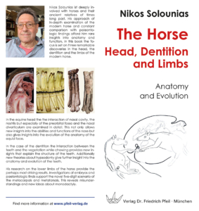
His most important contributions are the study of the Miocene Samos fauna, the development of tooth micro wear, the dietary evolution of horses through the Cenozoic, the explanation as to why some herbi­vores have tall (hypsodont) teeth and the discovery that the ruminants also have five digits in their foot.

Bewertungen

Es gibt noch keine Bewertungen.

Schreiben Sie die erste Bewertung für „The Horse – Head, Dentition and Limbs“ Antworten abbrechen

Sie müssen angemeldet sein, um eine Bewertung abgeben zu können.

„*“ zeigt erforderliche Felder an

Name*
E-Mail*
Anschrift*
Einwilligung*

Ähnliche Produkte

  • Fauna und Fazies der kondensierten Sedimente des Dogger und Malm (Bajocium bis Oxfordium) im südlichen Frankenjura

    30,00 €

    zzgl. Versandkosten / Versandkostenfrei in D

  • Die Stinkkalke der produktiven Cyrenenschichten Süddeutschlands zwischen Lech und Inn (Egerian)

    30,00 €

    zzgl. Versandkosten / Versandkostenfrei in D

  • Tertiäre Otolithen aus South-Australia und Victoria (Australien)

    20,00 €

    zzgl. Versandkosten / Versandkostenfrei in D

  • 6th International Meeting on Mesozoic Fishes

    20,00 €

    zzgl. Versandkosten / Versandkostenfrei in D

  • Spumellarienfaunen (Radiolaria) aus den mitteltriassischen Buchensteiner-Schichten von Recoaro (Norditalien) und den obertriassischen Reiflingerkalken von Großreifling (Österreich) – Systematik – Stratigraphie

    30,00 €

    zzgl. Versandkosten / Versandkostenfrei in D

Wir sind gerne für Sie da

Verlag Dr. Friedrich Pfeil
Wolfratshauser Str. 27
81379 Munich – Germany

Tel.: +49 89 55 28 6000
Fax: +49 89 55 28 6004
E-Mail: info@pfeil-verlag.de

Hauptstraße 12B - 85232 Bergkirchen OT Günding - Germany - Tel.: +49 8131 61 46 590 - Fax: +49 8131 61 46 591 - E-Mail: info@pfeil-verlag.de
© 2023 Verlag Dr. Friedrich Pfeil - Alle Rechte vorbehalten - Kontakt - Impressum - Datenschutzerklärung
Nach oben scrollen Nach oben scrollen Nach oben scrollen

Unsere Webseite verwendet erforderliche Cookies. Durch das Akzeptieren dieser Cookies sorgen Sie dafür, dass bei Ihrem Besuch alle Services sicher, stabil und reibungslos funktionieren. Die Verarbeitung Ihrer Daten erfolgt in Übereinstimmung mit unseren Datenschutz-Richtlinien

AkzeptierenNur essentielle CookiesEinstellungen

Cookies und Datenschutz-Einstellungen


Was sind Cookies?

Auf unserer Website verwenden wir sogenannte Cookies. Bei Cookies handelt es sich um Textdateien, die im Internetbrowser bzw. vom Internetbrowser auf dem Computersystem des Nutzers gespeichert werden, sobald ein Nutzer unsere Website aufruft. Cookies enthalten charakteristische Zeichenfolgen, die eine eindeutige Identifizierung des Browsers beim erneuten Aufrufen der Website ermöglichen.

Wir setzen Cookies ein, um unsere Website nutzerfreundlicher und sicher zu gestalten. Einige Seitenelemente unserer Website machen es erforderlich, dass der aufrufende Internetbrowser auch nach einem Seitenwechsel innerhalb unserer Website identifiziert werden kann.

Notwendige Cookies

Der Zweck der Verwendung technisch notwendiger Cookies ist es, die Nutzung von Websites für die Nutzer zu vereinfachen. Einige Funktionen unserer Internetseite können ohne den Einsatz von Cookies nicht angeboten werden. Für diese ist es erforderlich, dass der Internetbrowser auch nach einem Seitenwechsel wiedererkannt wird. In diesen technisch notwendigen Cookies werden Daten erhoben, gespeichert und an uns übermittelt, um den Abruf unserer Website aus der Datenbank zu ermöglichen.

Web Analytics

Wir setzen keinerlei fremde Dienste zur Analyse Ihres Surfverhaltens ein.

Unser Hostingprovider wertet von Zeit zu Zeit die erfolgten Zugriffe mithilfe von Werkzeugen aus, die auf die ohnehin anfallenden Serverlogfiles zugreifen. Diese Auswertung erfolgt ausschließlich anonymisiert und dient lediglich der Optimierung unserer Webdienste und der Server selbst.

Andere externe Dienste

Wir verwenden verschiedene externe Dienste wie z.B. Bayernatlas. Deren Anbieter sammeln möglicherweise personenbezogene Daten wie Ihre IP-Adresse.

Datenschutzerklärung

Wir bemühen uns, eine einfache und gut beschriebene Datenschutzerklärung bereitzustellen. Wir finden Datenschutz äußerst wichtig und wollen genau erklären, wie sorgsam wir mit Ihren Daten umgehen. Unsere Datenschutzerklärung finden Sie hier:

Datenschutzerklärung
AkzeptierenNur essentielle